Genetic testing of degraded embryos, FISH method for 5 chromosomes (13, 18, 21, X, Y) (including 10 samples).
Genetic testing of degraded embryos, FISH method for 5 chromosomes (13, 18, 21, X, Y) is a molecular cytogenetic study that allows to determine quantitative chromosomal abnormalities associated with the most common genetic disorders in the cells of an embryo that has stopped its development (degraded).
The FISH method is effective for detecting complex chromosomal aberrations and polyploidies, which is a characteristic feature of this type of preimplantation genetic testing. The FISH method is recommended for genetic diagnosis of embryos that have stopped in development. The information obtained will help to understand the potential cause of the developmental arrest and optimize further treatment with assisted reproductive technologies.
Testing includes 5 chromosomes to study quantitative abnormalities (changes in number) on chromosomes 13, 18, 21, X and Y.
Indications for the study Genetic testing of degraded embryos, FISH method of testing for 5 chromosomes (13, 18, 21, X, Y)
Detection during treatment with assisted reproductive technologies of embryos that have ceased to develop during the process of cultivation.
Why can embryos stop developing (degrade)?
- The presence of aneuploidies (a change in the number of chromosomes), such as monosomy (one chromosome missing) or trisomy (one additional chromosome) on one chromosome.
- Presence of complex aberrations (additional or missing several different chromosomes), polyploidies (increase or decrease in the number of chromosomes under study), incorrect distribution of chromosomes between cells.
- Age factors, the age of a woman over 38 years old. A decrease in the quality of eggs may indicate a decrease in the overall quality of the energy complex of eggs (mitochondria). Reduced quality of the energy complex leads to the accumulation of chromosomal changes that can cause embryo degradation.
- High level of sperm DNA fragmentation. During cultivation on the 3rd day of development, the so-called embryonic genome begins to function (only maternal DNA is involved before the 3rd day of development), so there may be disruptions in gene functioning, which leads to a stop in the growth and development of the embryo.
Advantages of the method
Genetic testing of degraded embryos will help to find out whether the cause of embryo developmental arrest is the presence of chromosomal abnormalities in the cells under study.
The information obtained will become a reliable source for further diagnosis and the formation of an individual treatment plan by a reproductive specialist.
Genetic testing of degraded embryos, FISH method for 5 chromosomes is recommended for patients who need analysis of degraded embryos, namely the determination of quantitative chromosomal abnormalities (abnormalities). The 5 chromosome screening panel includes the study of aneuploidies on chromosomes 13, 18, 21, X and Y.
The method is effective for detecting complex chromosomal aberrations (chromosome structure disorders) and polyploidies (an increase in the number of all the chromosomes under study) of embryos that have stopped development.
Preparation and course of the procedure
For genetic testing of degraded embryos, the FISH method is used.
FISH (fluorescence in situ hybridization) is a molecular cytogenetic method that helps to detect specific DNA sequences in the nuclei of cells using special reagents (fluorescently labeled probes):
- fixation of embryo cells on a glass slide.
- denaturation – separation of DNA strands under the influence of temperature, performed to allow the fluorescent reagent to bind to the DNA strand under study
- hybridization – the interaction of a fluorescent reagent with the DNA of the embryo under test
- sample purification to remove excess fluorescent reagents that have not bound to DNA
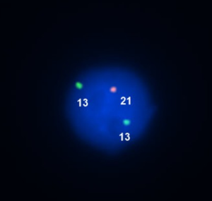
- The results are examined using a fluorescence microscope with a magnification of 1000 times. Specialists in the cytogenetic laboratory check for the presence of “control light signals”. Cells with a normal genetic set will show green, blue or orange glow in the appropriate amount of light signals.
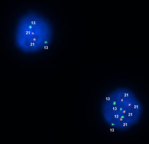
Visualization of the results of FISH analysis of cells from degraded embryos
Normal ( 2 orange signals and 2 green signals) (image in the upper left corner)
Polyploidy ( 4 orange and 4 green signals) (image in the lower right corner)
Aneuploidy
Aneuploidy, one 21 chromosome is missing